
¡¡Bienvenid@s a nuestra web!!
Iñaki González Restauración y Decoración es una empresa de nueva creación que surge tras el desarrollo, durante varios años, de estudios superiores en las áreas de la restauración y decoración artística. Nuestra labor abarca distintos campos de actuación dentro de la restauración: Tratamientos de madera, limpieza por chorreo, reintegración de elementos arquitectónicos, molduras, balaustradas etc... Además realizamos proyectos integrales de diseño y decoración de interiores y exteriores: Escultura, estucos, esgrafiados, imitaciones de piedra, ladrillo etc... Consulte nuestra gama de servicios y contacte con nosotros si necesita rehabilitar o redecorar su hogar o local comercial. Presupuestos sin compromiso.
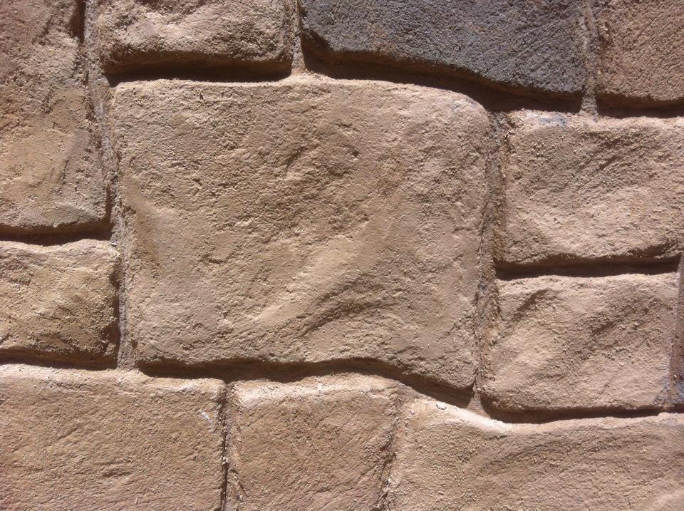
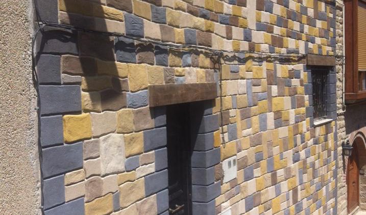
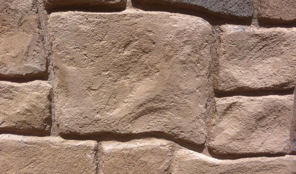
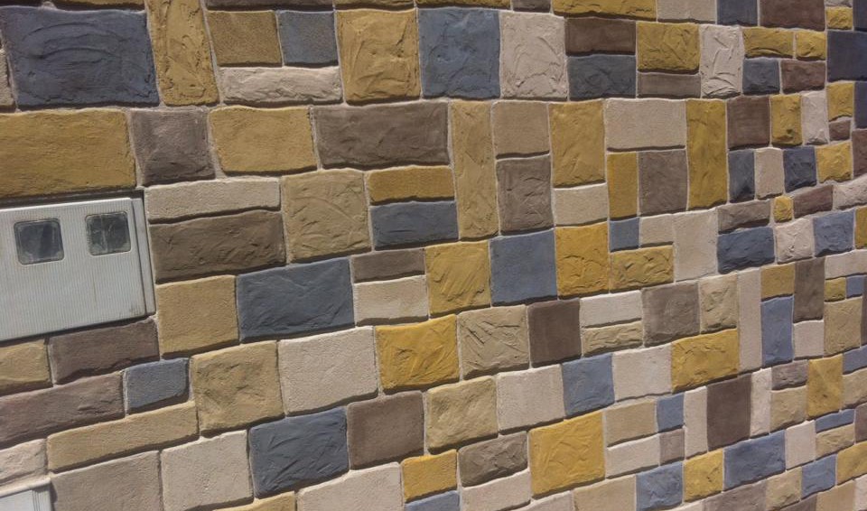
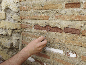
RESTAURACIÓN DE FACHADAS
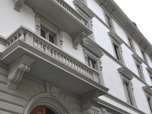
Restauración de fachadas e interiores de alta calidad. Conservación y rehabilitación de elementos estructurales. Chorreo por arena, limpieza química, alteraciones biológicas.
TRATAMIENTOS DE MADERA
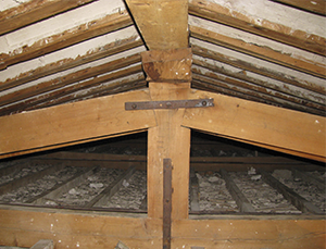
Tratamiento y rehabilitación de estructuras de madera. Tratamientos garantizados por un mínimo de 10 años contra todo tipo de xilófagos y agentes destructores de la madera.
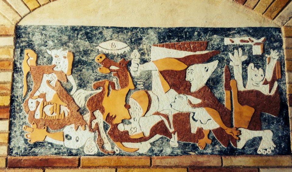
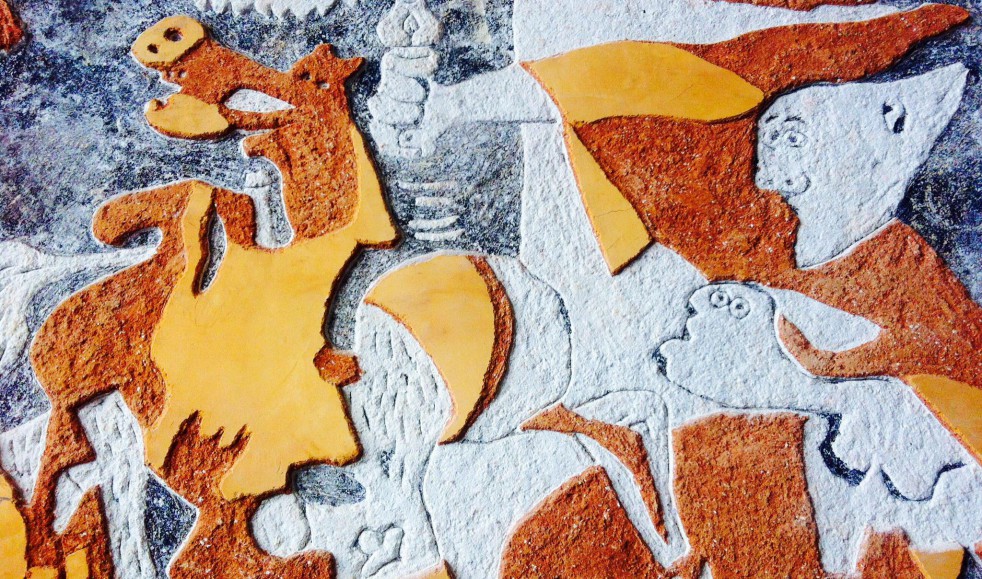
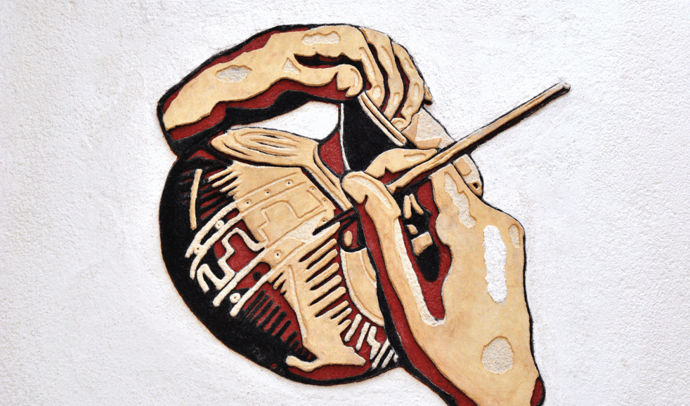
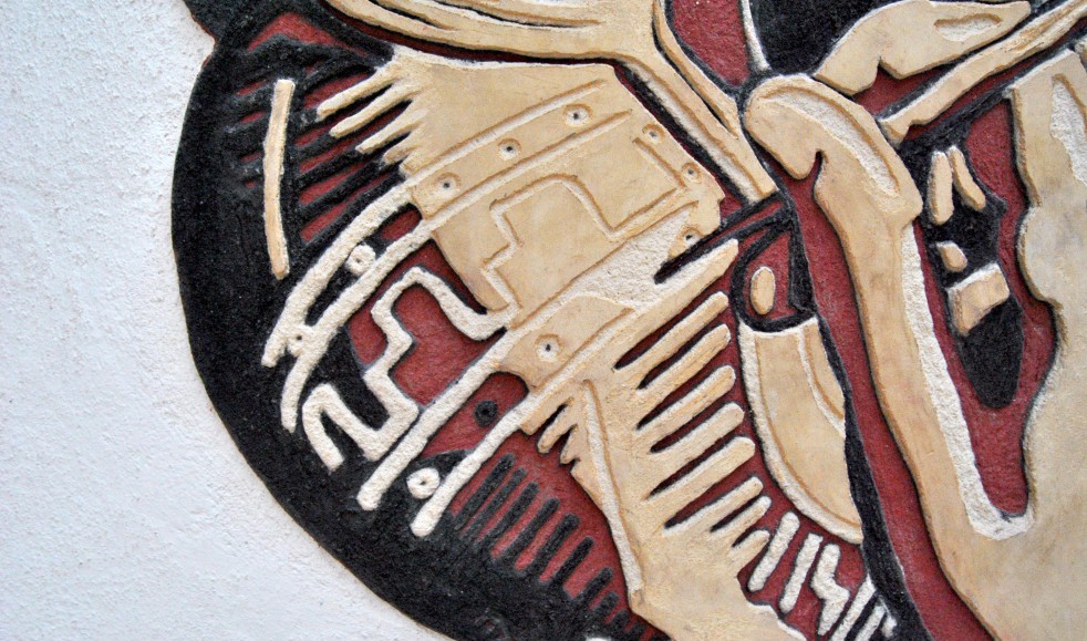
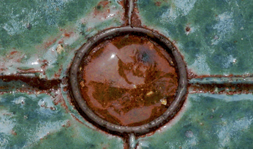
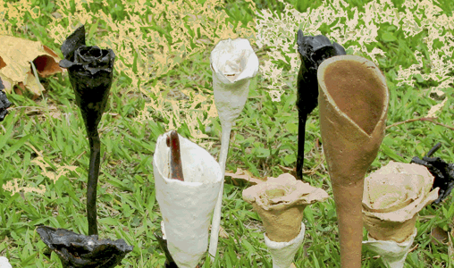
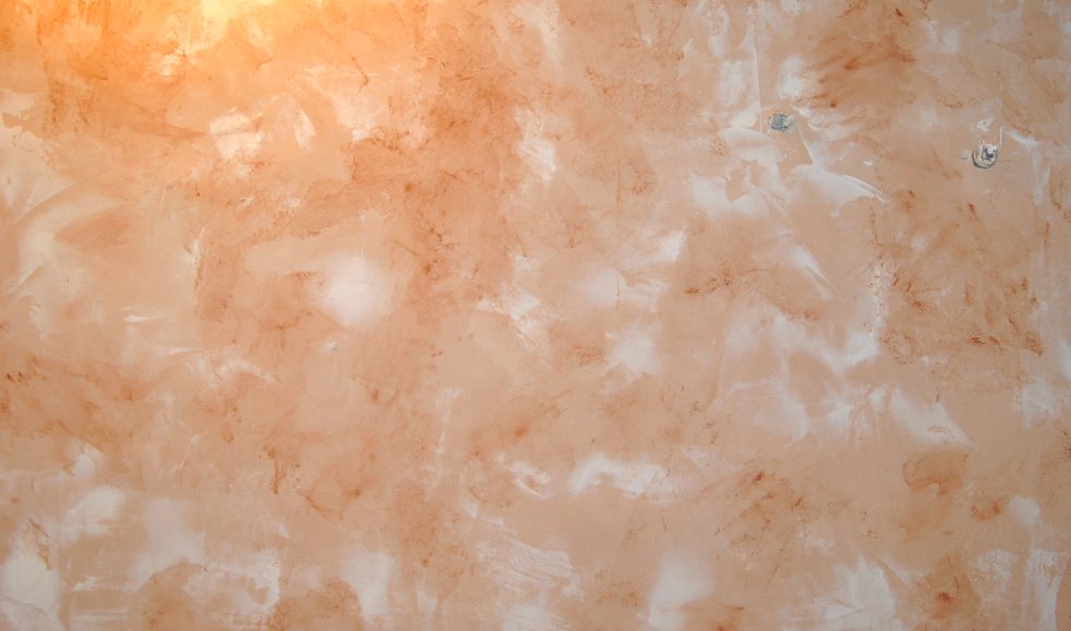
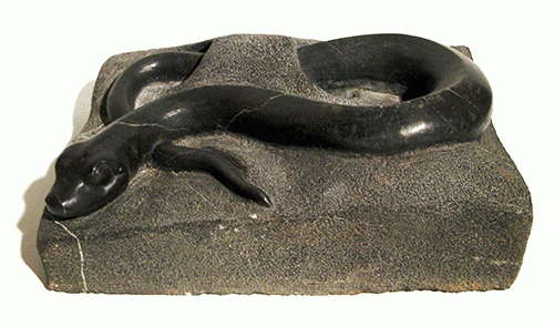
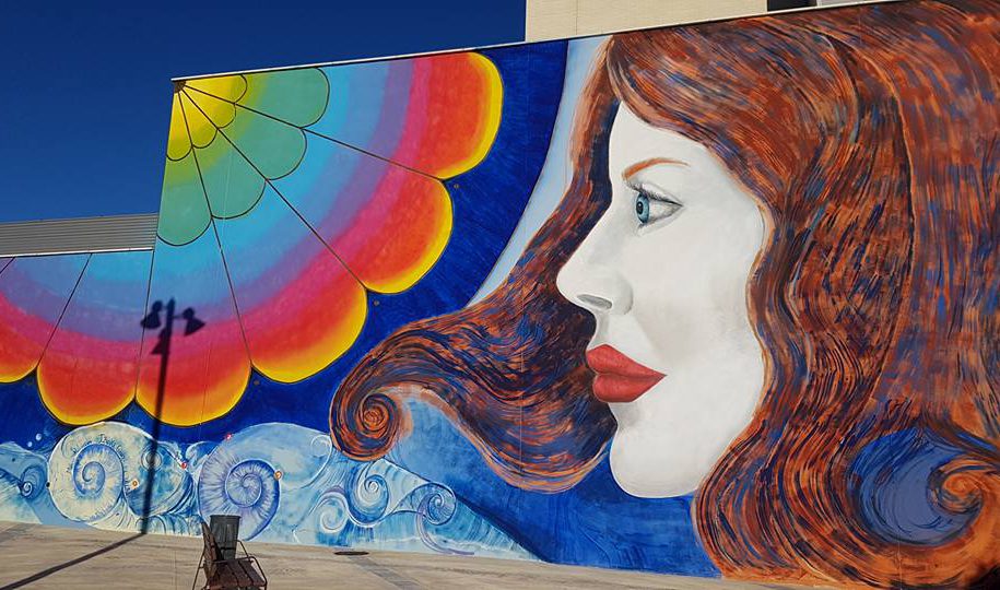
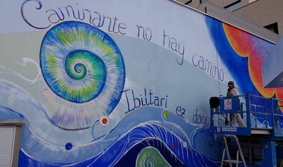
ALTA DECORACIÓN

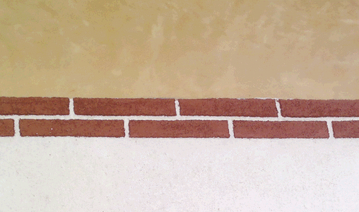
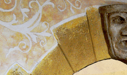
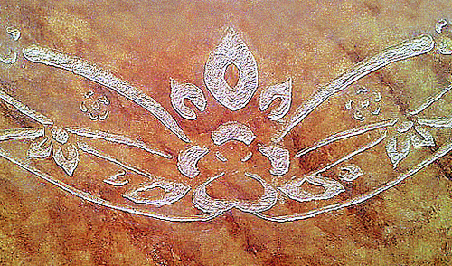
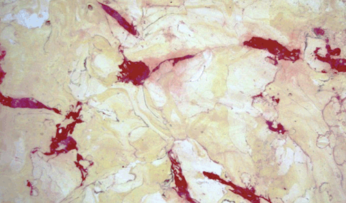
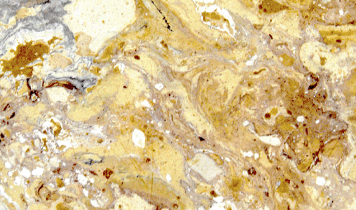
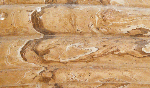
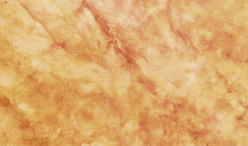
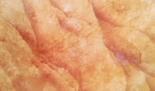
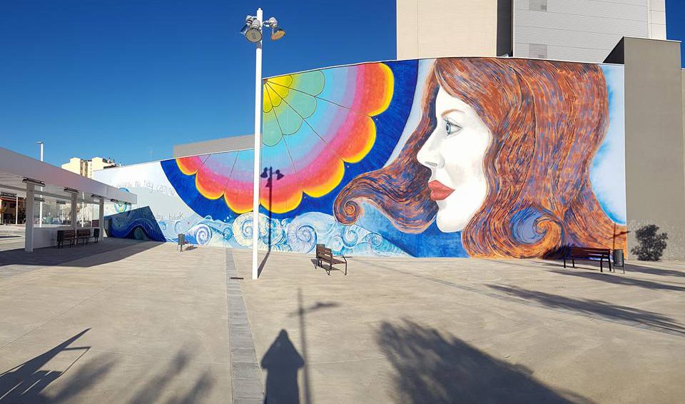
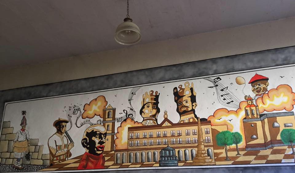
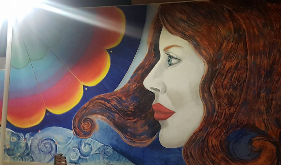
Alta decoración de exteriores e interiores. Pintura mural gran formato. Escultura, estucos, esgrafiados, imitaciones de mármol, cerámica artística...
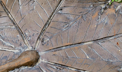
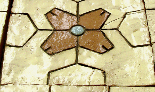
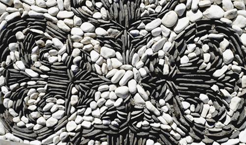
EMPEDRADO ARTÍSTICO
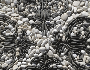
Restauración, conservación y decoración de empedrados artísticos granadinos. Dirección, gestión y realización de proyectos.
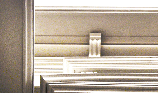
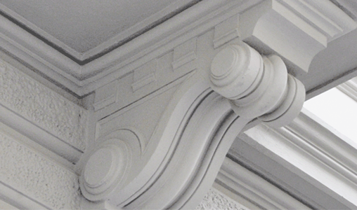
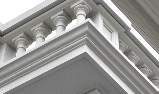
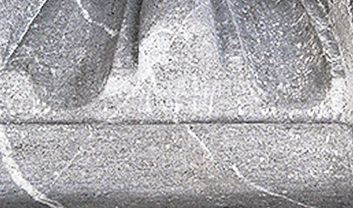
REINTEGRACIÓN
Reintegración de todo tipo de elementos arquitectónicos. Restauración de cornisas, molduras, balaustradas, escudos...
BIOCONSTRUCCIÓN